
User Manual Part 5

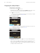
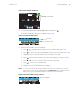
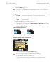


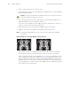
Table Of Contents

Directions for Use Standalone Monitoring 57
Monitoring ECG
Procedure
1. Inspect the ECG cable. Replace it if it shows any signs of wear, breakage, or fraying.
2. Plug the cable into the monitor.
3. Select electrode sites on the patient (Figure 54), choosing flat areas and avoiding fatty
or bony areas and major muscles.
Figure 54. ECG Leads - Actual Placement
Note
The monitor contains type CF fully isolated patient-connected circuitry, but it is
not intended for direct application on a patient’s heart.
Severe artifact and interference (such as defibrillation interference) can cause the
waveform to move off of the display for a few seconds before it is restored.
Impedance pneumography (Resp) is not recommended for use with
high-frequency ventilation.
The monitor counts as breaths any respiratory efforts larger than twice the
background cardiovascular artifact.
Use only silver/silver chloride electrodes. Other electrodes, such as stainless
steel electrodes, squeeze-bulb electrodes, or electrodes with dissimilar metals,
are subject to large offset potentials due to polarization. Other electrodes can also
have slower recovery time after the application of defibrillator pulses.
3-lead, adult and pediatric
3-lead, neonatal
5-lead, adult and pediatric
RA
LL
LA
RA
LL
LA
RA
LL
LA
V1
V6
RL
Six possible V lead electrode
placement sites for the C lead.