
Scanner User Manual
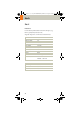
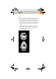
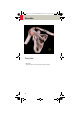
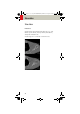
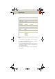
Table Of Contents
- User Documentation
- Scan and Reconstruction
- Dose Information
- Workflow Information
- Application Information
- Head
- Neck
- Shoulder
- Thorax
- Abdomen
- Pelvis
- Spine
- Upper Extremities
- Lower Extremities
- Vascular
- Specials
- Children
- Overview
- Hints in General
- HeadRoutine_Baby
- HeadRoutine_Child
- HeadSeq_Baby
- HeadSeq_Child
- InnerEar
- SinusOrbi
- Neck
- ThoraxRoutine_Baby
- ThoraxRoutine_Child
- ThoraxHRSeq_Baby
- ThoraxHRSeq_Child
- Abdomen_Baby
- Abdomen_Child
- Spine_Baby
- Spine_Child
- ExtrHR_Baby
- ExtrHR_Child
- HeadAngio
- HeadAngio08s
- CarotidAngio
- CarotidAngio08s
- BodyAngio
- BodyAngio08s
- NeonateBody
- syngo 3D
- syngo Fly Through
- syngo Dental CT
- syngo Osteo CT
- syngo Volume Evaluation
- syngo Dynamic Evaluation

Shoulder
97
Hints
• Use raw data to review a target region if necessary.
• For image reconstruction of soft tissue, use kernel
B31s and a slice width of 5.0 mm.
• Coronal and sagittal 2D planar reconstructions are
important for evaluation of the joint space & bursa
sacs in CT arthograms.
• 3D renderings are helpful for complex fractures &
dislocations.
Shoulder 2
nd
reconstruction
kV 130
Effective mAs 70
Rotation time 1.0 sec
Slice
collimation
1.5 mm
Slice width 3.0 mm 2.0 mm
Feed/Rotation 3.0 mm
Pitch Factor 1.0
Increment 3.0 mm 1.5 mm
Kernel B60s
CTDIVol
7.55 mGy
Effective dose Male:
0.94
mSv
Female:
1.13
mSv
C2-025.630.01.01.02_APPLICATIONGUIDE_SPIRIT.book Page 97 Friday, April 8, 2005 9:55 AM