
Scanner User Manual

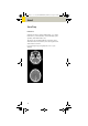
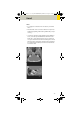
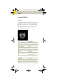
Table Of Contents
- User Documentation
- Scan and Reconstruction
- Dose Information
- Workflow Information
- Application Information
- Head
- Neck
- Shoulder
- Thorax
- Abdomen
- Pelvis
- Spine
- Upper Extremities
- Lower Extremities
- Vascular
- Specials
- Children
- Overview
- Hints in General
- HeadRoutine_Baby
- HeadRoutine_Child
- HeadSeq_Baby
- HeadSeq_Child
- InnerEar
- SinusOrbi
- Neck
- ThoraxRoutine_Baby
- ThoraxRoutine_Child
- ThoraxHRSeq_Baby
- ThoraxHRSeq_Child
- Abdomen_Baby
- Abdomen_Child
- Spine_Baby
- Spine_Child
- ExtrHR_Baby
- ExtrHR_Child
- HeadAngio
- HeadAngio08s
- CarotidAngio
- CarotidAngio08s
- BodyAngio
- BodyAngio08s
- NeonateBody
- syngo 3D
- syngo Fly Through
- syngo Dental CT
- syngo Osteo CT
- syngo Volume Evaluation
- syngo Dynamic Evaluation

Head
73
Hints
• An automatic bone correction allows for improved
head image quality, without any additional post-pro
-
cessing.
• In order to optimize image quality versus radiation
dose, scans are provided within a maximum scan
field of 300 mm with respect to the iso-center. No
recon job with a field of view exceeding those limits
will be possible. Therefore, patient positioning has to
be performed accurately to ensure a centered loca
-
tion of the skull.
Base Cerebrum
kV 130 130
Effective mAs 110 110
Rotation time 1.5 sec 1.5 sec
Slice collimation 1.5 mm 2.5 mm
Slice width 3.0 mm 8.0 mm
Feed/Rotation 3.0 mm 5.0 mm
Pitch Factor 1.0 1.0
Increment 3.0 mm 8.0 mm
Kernel H31s H31s
CTDIVol
25.05 mGy 25.05 mGy
Effective dose Male:
0.37
mSv
Female:
0.38
mSv
Male:
0.70
mSv
Female:
0.77
mSv
Contrast medium IV injection
Volume 50 – 60 ml
Flow rate 2 ml/sec.
Start delay 50 – 60 sec.
C2-025.630.01.01.02_APPLICATIONGUIDE_SPIRIT.book Page 73 Friday, April 8, 2005 9:55 AM