
Scanner User Manual


Table Of Contents
- User Documentation
- Scan and Reconstruction
- Dose Information
- Workflow Information
- Application Information
- Head
- Neck
- Shoulder
- Thorax
- Abdomen
- Pelvis
- Spine
- Upper Extremities
- Lower Extremities
- Vascular
- Specials
- Children
- Overview
- Hints in General
- HeadRoutine_Baby
- HeadRoutine_Child
- HeadSeq_Baby
- HeadSeq_Child
- InnerEar
- SinusOrbi
- Neck
- ThoraxRoutine_Baby
- ThoraxRoutine_Child
- ThoraxHRSeq_Baby
- ThoraxHRSeq_Child
- Abdomen_Baby
- Abdomen_Child
- Spine_Baby
- Spine_Child
- ExtrHR_Baby
- ExtrHR_Child
- HeadAngio
- HeadAngio08s
- CarotidAngio
- CarotidAngio08s
- BodyAngio
- BodyAngio08s
- NeonateBody
- syngo 3D
- syngo Fly Through
- syngo Dental CT
- syngo Osteo CT
- syngo Volume Evaluation
- syngo Dynamic Evaluation
314
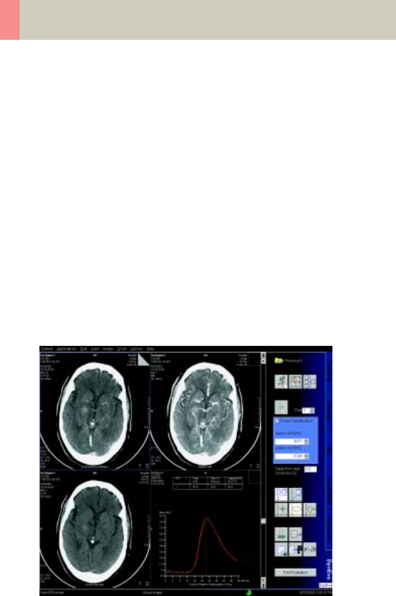
syngo Dynamic Evaluation
5. Evaluation of Region of Interests
You select the image regions to be evaluated by mark-
ing them with ROIs or applying the pixel lens (a circular
ROI with a fixed but configurable diameter). An abso-
lute/relative CT-value calculation is performed for
these selected image.
• You can draw either elliptical or freehand ROIs.
• You can modify an ROI in any image, precisely adapt-
ing it to the shape you feel is relevant.
• The number of ROIs that can be defined is limited to
five – you can only draw one pixel lens.
• The ROIs that you have drawn are transferred to the
other views.
• To aid examination, they are numbered in sequence
and color-coded.
Display of the Pixel Lens curve
C2-025.630.01.01.02_APPLICATIONGUIDE_SPIRIT.book Page 314 Friday, April 8, 2005 9:55 AM