
Scanner User Manual


Table Of Contents
- User Documentation
- Scan and Reconstruction
- Dose Information
- Workflow Information
- Application Information
- Head
- Neck
- Shoulder
- Thorax
- Abdomen
- Pelvis
- Spine
- Upper Extremities
- Lower Extremities
- Vascular
- Specials
- Children
- Overview
- Hints in General
- HeadRoutine_Baby
- HeadRoutine_Child
- HeadSeq_Baby
- HeadSeq_Child
- InnerEar
- SinusOrbi
- Neck
- ThoraxRoutine_Baby
- ThoraxRoutine_Child
- ThoraxHRSeq_Baby
- ThoraxHRSeq_Child
- Abdomen_Baby
- Abdomen_Child
- Spine_Baby
- Spine_Child
- ExtrHR_Baby
- ExtrHR_Child
- HeadAngio
- HeadAngio08s
- CarotidAngio
- CarotidAngio08s
- BodyAngio
- BodyAngio08s
- NeonateBody
- syngo 3D
- syngo Fly Through
- syngo Dental CT
- syngo Osteo CT
- syngo Volume Evaluation
- syngo Dynamic Evaluation
syngo Volume Evaluation
291
Workflow
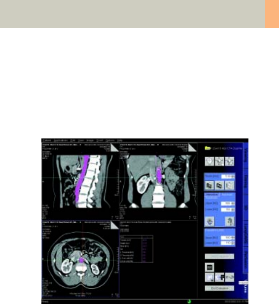
1. Loading the Images
After loading the images into syngo Volume Evalua-
tion, the following layout is displayed:
• Segment 1: Display of sagittal images.
The images are sorted from left to right, according to
the patient’s anatomy.
• Segment 2: Display of coronal images.
The images are sorted from front to back, according
to the patient’s anatomy.
• Segment 3: Display of transversal images.
The images are sorted from head to feet, according
to the patient’s anatomy.
• Segment 4: Display of either the evaluation results or
thick slice images (MaxIP, MinIP or MPR images) that
correspond to the transversal images in segment 3.
C2-025.630.01.01.02_APPLICATIONGUIDE_SPIRIT.book Page 291 Friday, April 8, 2005 9:55 AM