
Scanner User Manual
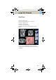

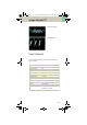
Table Of Contents
- User Documentation
- Scan and Reconstruction
- Dose Information
- Workflow Information
- Application Information
- Head
- Neck
- Shoulder
- Thorax
- Abdomen
- Pelvis
- Spine
- Upper Extremities
- Lower Extremities
- Vascular
- Specials
- Children
- Overview
- Hints in General
- HeadRoutine_Baby
- HeadRoutine_Child
- HeadSeq_Baby
- HeadSeq_Child
- InnerEar
- SinusOrbi
- Neck
- ThoraxRoutine_Baby
- ThoraxRoutine_Child
- ThoraxHRSeq_Baby
- ThoraxHRSeq_Child
- Abdomen_Baby
- Abdomen_Child
- Spine_Baby
- Spine_Child
- ExtrHR_Baby
- ExtrHR_Child
- HeadAngio
- HeadAngio08s
- CarotidAngio
- CarotidAngio08s
- BodyAngio
- BodyAngio08s
- NeonateBody
- syngo 3D
- syngo Fly Through
- syngo Dental CT
- syngo Osteo CT
- syngo Volume Evaluation
- syngo Dynamic Evaluation

syngo Fly Through
261
• Standing Point
– The cone rotates around the viewing point.
• Viewing Point
– The complete cone moves.
• Clipping Point
– The cone rotates around the standing point.
• Viewing Angle
– Is changed by moving one of the sides of the
angle.
• Viewing Distance
– The distance from the standing to the viewing
point.
• Viewing Depth
– Moving the Front/Back Clip Plane changes the
viewing depth.
• Front Clip Plane
– To remove foreground-obscuring tissue. The posi-
tion of the front clip plane will always be between
standing and viewing point.
•Back Clip Plane
– To remove tissue at the back of the volume. The
position of the back clip plane will always be
behind viewing point.
C2-025.630.01.01.02_APPLICATIONGUIDE_SPIRIT.book Page 261 Friday, April 8, 2005 9:55 AM