
Scanner User Manual
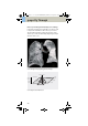
Table Of Contents
- User Documentation
- Scan and Reconstruction
- Dose Information
- Workflow Information
- Application Information
- Head
- Neck
- Shoulder
- Thorax
- Abdomen
- Pelvis
- Spine
- Upper Extremities
- Lower Extremities
- Vascular
- Specials
- Children
- Overview
- Hints in General
- HeadRoutine_Baby
- HeadRoutine_Child
- HeadSeq_Baby
- HeadSeq_Child
- InnerEar
- SinusOrbi
- Neck
- ThoraxRoutine_Baby
- ThoraxRoutine_Child
- ThoraxHRSeq_Baby
- ThoraxHRSeq_Child
- Abdomen_Baby
- Abdomen_Child
- Spine_Baby
- Spine_Child
- ExtrHR_Baby
- ExtrHR_Child
- HeadAngio
- HeadAngio08s
- CarotidAngio
- CarotidAngio08s
- BodyAngio
- BodyAngio08s
- NeonateBody
- syngo 3D
- syngo Fly Through
- syngo Dental CT
- syngo Osteo CT
- syngo Volume Evaluation
- syngo Dynamic Evaluation

syngo 3D
253
Using VRT/VRT Thin/Clip
1. Load the series to VRT.
2. Manipulate volume to evaluate anatomy:
a)Orientate the image volume for the best view of
the anatomy.
b)To eliminate unwanted bone, either activate VRT
Thin/Clip or use VOI punching/3D Object Editor.
c)Select VRT preset from VRT gallery.
d)Adjust window, opacity, color and lighting effects
as necessary.
e)Pan and zoom image as necessary.
3. Measure, annotate and document as needed.
4. Optionally, create image series using radial ranges
for VRT; parallel, expanded or curved ranges for VRT
Thin/Clip and output them either on a film or save
them to the database.
C2-025.630.01.01.02_APPLICATIONGUIDE_SPIRIT.book Page 253 Friday, April 8, 2005 9:55 AM