
Scanner User Manual


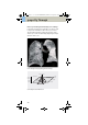
Table Of Contents
- User Documentation
- Scan and Reconstruction
- Dose Information
- Workflow Information
- Application Information
- Head
- Neck
- Shoulder
- Thorax
- Abdomen
- Pelvis
- Spine
- Upper Extremities
- Lower Extremities
- Vascular
- Specials
- Children
- Overview
- Hints in General
- HeadRoutine_Baby
- HeadRoutine_Child
- HeadSeq_Baby
- HeadSeq_Child
- InnerEar
- SinusOrbi
- Neck
- ThoraxRoutine_Baby
- ThoraxRoutine_Child
- ThoraxHRSeq_Baby
- ThoraxHRSeq_Child
- Abdomen_Baby
- Abdomen_Child
- Spine_Baby
- Spine_Child
- ExtrHR_Baby
- ExtrHR_Child
- HeadAngio
- HeadAngio08s
- CarotidAngio
- CarotidAngio08s
- BodyAngio
- BodyAngio08s
- NeonateBody
- syngo 3D
- syngo Fly Through
- syngo Dental CT
- syngo Osteo CT
- syngo Volume Evaluation
- syngo Dynamic Evaluation

252
syngo 3D
Workflow for a CT
Angiography
Using MIP/MIP Thin
1. Load the series to MIP.
2. Manipulate the volume to evaluate anatomy:
a)Orientate the image volume for the best view of
the anatomy.
b)To eliminate unwanted bone, either activate MIP
Thin or use VOI punching/3D Object Editor.
c)Adjust the window as necessary.
d)Pan and zoom image as necessary.
3. Measure, annotate and document as needed.
4. Optionally, create image series using radial ranges
for MIP; parallel, expanded or curved ranges for MIP
Thin and output them either on a film or save them
to the database.
C2-025.630.01.01.02_APPLICATIONGUIDE_SPIRIT.book Page 252 Friday, April 8, 2005 9:55 AM