
Scanner User Manual


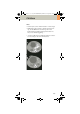

Table Of Contents
- User Documentation
- Scan and Reconstruction
- Dose Information
- Workflow Information
- Application Information
- Head
- Neck
- Shoulder
- Thorax
- Abdomen
- Pelvis
- Spine
- Upper Extremities
- Lower Extremities
- Vascular
- Specials
- Children
- Overview
- Hints in General
- HeadRoutine_Baby
- HeadRoutine_Child
- HeadSeq_Baby
- HeadSeq_Child
- InnerEar
- SinusOrbi
- Neck
- ThoraxRoutine_Baby
- ThoraxRoutine_Child
- ThoraxHRSeq_Baby
- ThoraxHRSeq_Child
- Abdomen_Baby
- Abdomen_Child
- Spine_Baby
- Spine_Child
- ExtrHR_Baby
- ExtrHR_Child
- HeadAngio
- HeadAngio08s
- CarotidAngio
- CarotidAngio08s
- BodyAngio
- BodyAngio08s
- NeonateBody
- syngo 3D
- syngo Fly Through
- syngo Dental CT
- syngo Osteo CT
- syngo Volume Evaluation
- syngo Dynamic Evaluation

240
syngo 3D
The 3D card offers the possibility to combine two-
dimensional images to form three-dimensional views.
To do this, the 3D card provides you with the following
methods:
Multi Planar Reconstruction (MPR)
Interactive navigation through 3D volumes in arbitrary
orientations.
• Orthogonal, oblique or double-oblique orientation.
• Easy scrolling through 3D volume data set.
• Real-time reconstruction of secondary slices.
• Additional diagnostic information, e.g., with sagittal
or coronal reconstructions from axial images.
• Variable slice thickness (MPR Thick, MPR) and dis-
tance with configurable default values.
• Calculation of arbitrarily curved cuts.
Maximum Intensity Projection (MIP)
Angiographic display.
• Projection of pixels with highest intensity (vascular
information) onto an arbitrarily oriented plane.
• Display of e.g., aneurysms, plaques, stenoses and
other vascular anomalies.
• MIP Thin function for projection within a small slab
to focus on particular vascular structure.
C2-025.630.01.01.02_APPLICATIONGUIDE_SPIRIT.book Page 240 Friday, April 8, 2005 9:55 AM