
Scanner User Manual

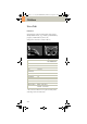
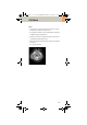
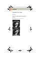

Table Of Contents
- User Documentation
- Scan and Reconstruction
- Dose Information
- Workflow Information
- Application Information
- Head
- Neck
- Shoulder
- Thorax
- Abdomen
- Pelvis
- Spine
- Upper Extremities
- Lower Extremities
- Vascular
- Specials
- Children
- Overview
- Hints in General
- HeadRoutine_Baby
- HeadRoutine_Child
- HeadSeq_Baby
- HeadSeq_Child
- InnerEar
- SinusOrbi
- Neck
- ThoraxRoutine_Baby
- ThoraxRoutine_Child
- ThoraxHRSeq_Baby
- ThoraxHRSeq_Child
- Abdomen_Baby
- Abdomen_Child
- Spine_Baby
- Spine_Child
- ExtrHR_Baby
- ExtrHR_Child
- HeadAngio
- HeadAngio08s
- CarotidAngio
- CarotidAngio08s
- BodyAngio
- BodyAngio08s
- NeonateBody
- syngo 3D
- syngo Fly Through
- syngo Dental CT
- syngo Osteo CT
- syngo Volume Evaluation
- syngo Dynamic Evaluation

Children
207
* The conversion factor for an 8-week-old child, and a
scan range of 150 mm was used.
Hints
• Use this protocol for children below 35 kg.
• The first and second recon jobs are defined for visu-
alization of the mediastinum and the lungs, respec-
tively.
• Please change the mAs value according to the
weight of the child:
25 – 34 kg 60 mAs.
ThorRoutine 2
nd
reconstruction
kV 80
Effective mAs 41
Rotation time 1.0 sec
Slice
collimation
2.5 mm
Slice width 5.0 mm 5.0 mm
Feed/Rotation 10.0 mm
Pitch Factor 2.0
Increment 5.0 mm 5.0 mm
Kernel B41s B60s
CTDIVol 1.13 mGy
Effective dose Male: 1.55 mSv*
Female: 1.85 mSv*
Contrast medium IV injection
Start delay exam dependent
Flow rate dependent upon needle size/
Access site
Total amount 1 – 2 ml per kg of body weight
C2-025.630.01.01.02_APPLICATIONGUIDE_SPIRIT.book Page 207 Friday, April 8, 2005 9:55 AM