
Scanner User Manual

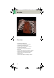


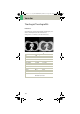

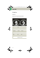
Table Of Contents
- User Documentation
- Scan and Reconstruction
- Dose Information
- Workflow Information
- Application Information
- Head
- Neck
- Shoulder
- Thorax
- Abdomen
- Pelvis
- Spine
- Upper Extremities
- Lower Extremities
- Vascular
- Specials
- Children
- Overview
- Hints in General
- HeadRoutine_Baby
- HeadRoutine_Child
- HeadSeq_Baby
- HeadSeq_Child
- InnerEar
- SinusOrbi
- Neck
- ThoraxRoutine_Baby
- ThoraxRoutine_Child
- ThoraxHRSeq_Baby
- ThoraxHRSeq_Child
- Abdomen_Baby
- Abdomen_Child
- Spine_Baby
- Spine_Child
- ExtrHR_Baby
- ExtrHR_Child
- HeadAngio
- HeadAngio08s
- CarotidAngio
- CarotidAngio08s
- BodyAngio
- BodyAngio08s
- NeonateBody
- syngo 3D
- syngo Fly Through
- syngo Dental CT
- syngo Osteo CT
- syngo Volume Evaluation
- syngo Dynamic Evaluation

Vascular
163
Hints in General
• Topogram: TOP, 512/1024 or LAT 256
• Patient positioning:
Patient lying in supine position, arms positioned
comfortably above the head in the head-arm rest,
lower legs supported.
• Patient respiratory instructions: inspiration.
• Oral administration of contrast medium:
The use of water will not obscure the blood vessels,
thus allowing CTA post-processing to be performed
easily afterwards.
• Be careful when examining pheochromocytoma
patients. Administration of an IV CM injection in
such cases may trigger a hypertensive crisis!
• To further optimize MPR image quality, we recom-
mend that you reduce one or more of the following
parameters: collimation, reconstruction increment
and slice width for image reconstruction.
Head Kernels
• For soft tissue head studies, the standard kernel is
H41s; softer images are obtained with H31s or H21s,
sharper images with H50s.
Body Kernels
• As standard kernels for body tissue studies B31s or
B41s is recommended; softer images are obtained
with B20s.
C2-025.630.01.01.02_APPLICATIONGUIDE_SPIRIT.book Page 163 Friday, April 8, 2005 9:55 AM