
Scanner User Manual
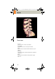

Table Of Contents
- User Documentation
- Scan and Reconstruction
- Dose Information
- Workflow Information
- Application Information
- Head
- Neck
- Shoulder
- Thorax
- Abdomen
- Pelvis
- Spine
- Upper Extremities
- Lower Extremities
- Vascular
- Specials
- Children
- Overview
- Hints in General
- HeadRoutine_Baby
- HeadRoutine_Child
- HeadSeq_Baby
- HeadSeq_Child
- InnerEar
- SinusOrbi
- Neck
- ThoraxRoutine_Baby
- ThoraxRoutine_Child
- ThoraxHRSeq_Baby
- ThoraxHRSeq_Child
- Abdomen_Baby
- Abdomen_Child
- Spine_Baby
- Spine_Child
- ExtrHR_Baby
- ExtrHR_Child
- HeadAngio
- HeadAngio08s
- CarotidAngio
- CarotidAngio08s
- BodyAngio
- BodyAngio08s
- NeonateBody
- syngo 3D
- syngo Fly Through
- syngo Dental CT
- syngo Osteo CT
- syngo Volume Evaluation
- syngo Dynamic Evaluation

134
Spine
• The CT scan following myelography must be per-
formed within 4 – 6 hours of the injection, other-
wise, the contrast density in the spinal canal will be
too high to obtain artifact-free images. Also, if possi
-
ble, it is a good idea to roll the patient once, or scan
in a prone position. This will prevent the contrast
from pooling posterior to the spinal cord.
• If a prone scan is performed, breathing instructions
are recommended to avoid motion artifact in axial
source and MPR images.
• To further optimize MPR image quality, we recom-
mend that you reduce one or more of the following
parameters: collimation, reconstruction increment,
and slice width for image reconstruction.
C2-025.630.01.01.02_APPLICATIONGUIDE_SPIRIT.book Page 134 Friday, April 8, 2005 9:55 AM