
Scanner User Manual

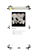


Table Of Contents
- User Documentation
- Scan and Reconstruction
- Dose Information
- Workflow Information
- Application Information
- Head
- Neck
- Shoulder
- Thorax
- Abdomen
- Pelvis
- Spine
- Upper Extremities
- Lower Extremities
- Vascular
- Specials
- Children
- Overview
- Hints in General
- HeadRoutine_Baby
- HeadRoutine_Child
- HeadSeq_Baby
- HeadSeq_Child
- InnerEar
- SinusOrbi
- Neck
- ThoraxRoutine_Baby
- ThoraxRoutine_Child
- ThoraxHRSeq_Baby
- ThoraxHRSeq_Child
- Abdomen_Baby
- Abdomen_Child
- Spine_Baby
- Spine_Child
- ExtrHR_Baby
- ExtrHR_Child
- HeadAngio
- HeadAngio08s
- CarotidAngio
- CarotidAngio08s
- BodyAngio
- BodyAngio08s
- NeonateBody
- syngo 3D
- syngo Fly Through
- syngo Dental CT
- syngo Osteo CT
- syngo Volume Evaluation
- syngo Dynamic Evaluation

Pelvis
125
Hints in General
• Topogram: TOP, 512 mm for pelvis studies and
256 mm for studies of the hip and SI Joints.
• Patient positioning:
Patient lying in supine position, arms positioned
comfortably above the head in the head-arm rest,
lower legs supported.
• A breathing command is not necessarily required for
the pelvic examination, since respiration does not
negatively influence this region.
• Rectal contrast medium administration:
Rectal contrast media is usually required to delineate
the rectum and sigmoid colon, if lower pelvic mass
or pathology are suspected. In some cases, air may
be substituted for a positive contrast agent. The use
of a vaginal tampon may be helpful in adult female
patients with suspected pelvis pathology.
• To further optimize MPR image quality we recom-
mend that you reduce one or more of the following:
collimation, reconstruction increment, and slice
width for image reconstruction.
• For pelvis studies, deselect CARE Dose for patients
>
120 kg.
Body Kernels
• As standard kernels for body tissue studies B31s or
B41s is recommended; softer images are obtained
with B20s.
C2-025.630.01.01.02_APPLICATIONGUIDE_SPIRIT.book Page 125 Friday, April 8, 2005 9:55 AM