
Scanner User Manual
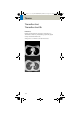


Table Of Contents
- User Documentation
- Scan and Reconstruction
- Dose Information
- Workflow Information
- Application Information
- Head
- Neck
- Shoulder
- Thorax
- Abdomen
- Pelvis
- Spine
- Upper Extremities
- Lower Extremities
- Vascular
- Specials
- Children
- Overview
- Hints in General
- HeadRoutine_Baby
- HeadRoutine_Child
- HeadSeq_Baby
- HeadSeq_Child
- InnerEar
- SinusOrbi
- Neck
- ThoraxRoutine_Baby
- ThoraxRoutine_Child
- ThoraxHRSeq_Baby
- ThoraxHRSeq_Child
- Abdomen_Baby
- Abdomen_Child
- Spine_Baby
- Spine_Child
- ExtrHR_Baby
- ExtrHR_Child
- HeadAngio
- HeadAngio08s
- CarotidAngio
- CarotidAngio08s
- BodyAngio
- BodyAngio08s
- NeonateBody
- syngo 3D
- syngo Fly Through
- syngo Dental CT
- syngo Osteo CT
- syngo Volume Evaluation
- syngo Dynamic Evaluation
Thorax
109
Hints
• You could repeat the same protocol by simply click-
ing the chronicle with the right mouse button for
“repeat“, e.g., when both non-contrast and contrast
studies are required.
• For lung cancer evaluation, this protocol can be com-
bined with the protocol “Neck Routine”.
• Low dose lung images are usually evaluated using
lung window setting. Soft tissue/bone window set
-
tings may be used to visualize the presence of calci-
fications in the nodules.
• It is essential to use the same protocol for follow-up
studies to check for progression.
LungLowDose
kV 130
Effective mAs 30
Rotation time 1.0 sec
Slice collimation 5.0 mm
Slice width 8.0 mm
Feed/Rotation 10.0 mm
Pitch Factor 1.0
Increment 8.0 mm
Kernel B70s
CTDIVol
3.23 mGy
Effective dose Male: 1.69 mSv
Female: 2.08 mSv
Contrast medium IV injection
Start delay 30 sec.
Flow rate 2.5 ml/sec.
Total amount 50 – 70 ml
C2-025.630.01.01.02_APPLICATIONGUIDE_SPIRIT.book Page 109 Friday, April 8, 2005 9:55 AM