
Scanner User Manual
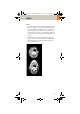
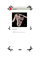
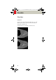
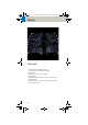
Table Of Contents
- User Documentation
- Scan and Reconstruction
- Dose Information
- Workflow Information
- Application Information
- Head
- Neck
- Shoulder
- Thorax
- Abdomen
- Pelvis
- Spine
- Upper Extremities
- Lower Extremities
- Vascular
- Specials
- Children
- Overview
- Hints in General
- HeadRoutine_Baby
- HeadRoutine_Child
- HeadSeq_Baby
- HeadSeq_Child
- InnerEar
- SinusOrbi
- Neck
- ThoraxRoutine_Baby
- ThoraxRoutine_Child
- ThoraxHRSeq_Baby
- ThoraxHRSeq_Child
- Abdomen_Baby
- Abdomen_Child
- Spine_Baby
- Spine_Child
- ExtrHR_Baby
- ExtrHR_Child
- HeadAngio
- HeadAngio08s
- CarotidAngio
- CarotidAngio08s
- BodyAngio
- BodyAngio08s
- NeonateBody
- syngo 3D
- syngo Fly Through
- syngo Dental CT
- syngo Osteo CT
- syngo Volume Evaluation
- syngo Dynamic Evaluation

100
Thorax
• Lung images should be documented in both soft tis-
sue window and lung window.
• It is also possible to interleave the soft tissue & lung
setting images in one film sheet. This can be set up
in the configuration for filming.
• To further optimize MPR image quality, we recom-
mend that you reduce one or more of the following
parameters: collimation, reconstruction increment,
and slice width for image reconstruction.
C2-025.630.01.01.02_APPLICATIONGUIDE_SPIRIT.book Page 100 Friday, April 8, 2005 9:55 AM