
Specifications

7Principles of Operation
2. Tidal Volume Adjustment
Inspiratory (V
T
I
) and expiratory (V
T
E
) tidal volumes are determined by the estimated patient flow,
and compared on a breath-by-breath basis. If the measured volumes during inspiration differ from
expiration, the difference in volume is assumed to be due to an unintentional circuit leak. The baseline
(V
leak
) is adjusted in the appropriate direction to reduce the difference in V
T
I
- V
T
E
on the next breath.
This prevents abrupt changes in sensitivity based on random changes in the breathing pattern, and
allows the baseline (V
leak
) to accommodate to the new breathing pattern.
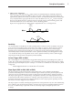
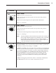
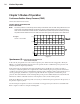
Figure 4 shows graphically how tidal volume is adjusted in the case of a change in leak.
V
est
V
T
0
0
Additional
Leak
Introduced
Volume
Adjustment
Figure 4: Tidal Volume Adjustment
•
•
Sensitivity
An essential feature of the BiPAP S/T while operating in the S and S/T modes is its ability to effectively
sense spontaneous breathing efforts, which causes the ventilator to trigger to IPAP and cycle to EPAP.
Because no preset sensitivity threshold can ensure patient and machine synchrony with changing
breathing efforts and circuit leaks, the BiPAP S/T continuously tracks patient breathing patterns and
automatically adjusts sensitivity thresholds to ensure optimum sensitivity as breathing patterns change
or as circuit leaks change. The algorithms used to ensure optimum sensitivity are the Volume Trigger,
Shape Signal, and the Spontaneous Expiratory Threshold (SET).
Volume Trigger (EPAP to IPAP)
The volume trigger is one method used to trigger IPAP during spontaneous breathing in the S and S/T
modes. The volume trigger threshold is 6 cc of accumulated volume above the baseline leak (V
leak
). When
patient effort generates inspiratory flow causing 6 cc of volume to accumulate above baseline (V
leak
), IPAP
is triggered:
Volume trigger threshold = 6 cc volume above V
leak
baseline
•
•
•
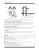
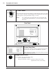
Shape Signal (EPAP to IPAP; IPAP to EPAP)
The shape signal is another method used to trigger IPAP and/or cycle off IPAP to EPAP during
spontaneous breathing in the S and S/T modes. This signal continuously tracks patient inspiratory and
expiratory flow rate and adjusts the spontaneous trigger and cycle thresholds for optimum sensitivity.
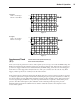
The shape signal appears as a shadow image of the patient's actual flow. The shape signal functions as a
sensitivity threshold at either inspiration or expiration. When the patient's flow rate crosses the shape
signal, the unit changes pressure levels. Figure 5 illustrates how the shape signal is superimposed onto
the actual waveform to trigger and cycle off IPAP.
The shape signal is created by offsetting the signal from the actual patient flow by 15 L/min and delaying
it for a 300 msec period. This intentional delay causes the shape signal to be slightly behind the patient's
flow rate. A sudden change in patient flow will cross the shape signal, causing the pressure level
to change.