
Operator's Manual
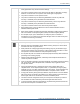
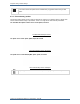
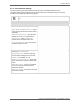
Table Of Contents

6.3 Pulse oximetry
Tempus Pro User/Operator Manual - 41-2001EN-00 – Page 97
6.3.4 Features
Depending on which features have been purchased for your Masimo Rainbow Pulse OX and which sensor
you are using will depend on which of the following features the Tempus Pro can monitor.
Masimo
®
SET
®
measurements supplied as standard:
SpO
2
- Intended for non-invasive measurement of arterial blood saturation and also provides pulse rate
when ECG is not attached.
Pulse Rate (PR).
Perfusion Index (PI) - Gives a numerical indication of the level of arterial pulsatile blood at the sensor
site.
Masimo
®
SET
®
Rainbow measurements purchased as optional upgrades:
SpCO - Allows clinicians to noninvasively and immediately detect elevated levels of carbon monoxide.
SpMet - Allows clinicians to noninvasively and immediately detect elevated levels of methaemoglobin in
the blood.
SpHb Index - Gives continuous monitoring of the haemoglobin levels in blood.
SpOC - Gives the patient’s oxygenation status by calculating the haemoglobin and oxygen saturation.
PVI purchased as an optional upgrade:
Pleth Variability Index (PVI) - Helps clinicians noninvasively and continuously assess fluid status of
patients.
The Signal Quality waveform (underneath the plethysmogram) shows how well the pulse sensor is detecting
the pulse. The height of the peak indicates the strength of the signal, if the peaks are short the finger sensor
should be repositioned.
The Pulse Rate displayed on the Tempus Pulse Oximeter may differ slightly from the heart rate displayed on
ECG monitors due to differences in averaging times. There may also be a discrepancy between cardiac
electrical activity and peripheral arterial pulsation. Significant differences may indicate a problem with the
signal quality due to physiological changes in the patient or one of the instruments or application of the
sensor or patient cable. The pulsations from intra-aortic balloon support can cause the pulse rate displayed
on the Tempus to be significantly different than the ECG heart rate.
The device indicates perfusion. It has been suggested that at extremely low perfusion levels, pulse
oximeters can measure peripheral saturation, which may differ from central arterial saturation. This
“localized hypoxemia” may result from the metabolic demands of other tissues extracting oxygen proximal to
the monitoring site under conditions of sustained peripheral hypoperfusion. (This may occur even with a
pulse rate that correlates with the ECG heart rate).
The SpCO index, Perfusion index
and Pleth Variability Index of the
site are shown here
The Pulse Oximetry section of the home screen
The
plethysmogram and
signal strength are
shown continuously
Masimo Rainbow parameters are
displayed here
The SpO
2
reading is
shown here