
Data Sheet

Electroencephalography (EEG)
Sensor Data Sheet
EEG
2601
20
18
PLUX – Wireless Biosignals, S.A.
Av. 5 de Outubro, n. 70 – 8.
1050-059 Lisbon, Portugal
plux@plux.info
http://biosignalsplux.com/
REV A
© 2015 PLUX
This information is provided "as is," and we make no express or implied warranties whatsoever with respect to functionality, operability, use,
fitness for a particular purpose, or infringement of rights. We expressly disclaim any liability whatsoever for any direct, indirect, consequential,
incidental or special damages, including, without limitation, lost revenues, lost profits, losses resulting from business interruption or loss of data,
regardless of the form of action or legal the
ory under which the liability may be asserted, even if advised of the possibility of such damages
.
SPECIFICATIONS
> Gain: 40000
> Range: ±37.5μV (with VCC = 3V)
> Bandwidth: 0.8-49Hz
> Consumption: ~3mA
> Input Impedance: >100GOhm
> CMRR: 100dB
FEATURES
> Single-channel differential sensor
> Discrete elastic head band
> Pre-conditioned analog output
> High signal-to-noise ratio
> Shielded miniaturized cables
> Medical-grade raw data output
> Ready-to-use form factor
APPLICATIONS
> Evoked potentials analysis
> Neurofeedback
> Sleep studies
> Human-Computer Interaction
> Neurophysiology studies
> Psychophysiology
GENERAL DESCRIPTION
Our electroencephalography (EEG) sensor
has been especially designed for both classic
and localized EEG measurement. When a
cap is too intrusive, only a limited number of
channels are needed, or you’d like to
synchronously record EEG and non-EEG
biosignals, this is the perfect solution. The
bipolar configuration, with two measurement
electrodes detects the electrical potentials in
the specific scalp region with respect to a
reference electrode, which should be placed
in a region of low muscular activity. The
resulting signal is the amplified difference
between these two signals, eliminating the
common unwanted signals detected by the
surfaces. Its convenient form factor enables a
discrete placement in regions such as the
forehead, occipital, and others. Examples:
http://bit.ly/1E7VenV
http://bit.ly/1PEskAZ
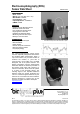
Fig. 1. The sensor is provided with a convenient elastic
head band to help secure the electrodes in place.
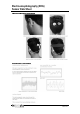
Fig. 2. Typical raw EEG data (acquired with biosignals).
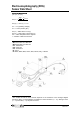
Fig. 3. Example
sensor
placemen
t
for localized EEG
.