
Use Instructions


PN 10232 Eagle V1.2 Instructions for Use Rev 1.0 Page 9 of 101
amino-4-oxo-pentanoic acid hydrochloride) for oral solution. When reconstituted in water, it can be
administered exogenously to overload the cellular porphyrin metabolism and generate a build-up of PpIX.
When ALA HCl is administered exogenously by oral route, the abundantly produced red fluorescent PpIX
cannot be quickly converted to its final product, heme, by the enzyme ferrochelatase and therefore
accumulates in cells. PpIX accumulates in cancer cells to a higher extent than in healthy cells. This is
primarily attributed to differences in the overall metabolic activity, increased ALA uptake, and a relative
deficiency in activity of ferrochelatase and other heme biosynthesis enzymes in cancer cells. The
preferential and selective accumulation of PpIX in cancer cells, relative to healthy cells, provides an
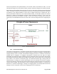
opportunity to visualize PpIX fluorescence in cancer cells in real-time during breast cancer surgery (Figure
1).
Figure 1 – Principles of Fluorescence
1.4.2. Fluorescence Imaging
The Eagle V1.2 Imaging System is a handheld intraoperative white light and Fluorescence Imaging device
and accessories intended to be used during surgical procedures. Fluorescence imaging is a type of optical
imaging technique used to visualize and document biological processes or structures. Fluorescence
imaging relies on exogenous or endogenous fluorophores, molecules that emit light of a longer
wavelength when exposed to light of a different (shorter) wavelength. When a fluorophore absorbs light,
the energy of the molecule is briefly raised to higher excited states. The subsequent return to the ground
state results in the emission of fluorescent light. The emitted fluorescent light, resulting from the
absorbed photon of energy has a specific wavelength and can be detected and measured. Typically,
Fluorescence Imaging is performed using steady-state fluorescence, where fluorophores are excited by a
constant source of light, emit fluorescence, and the emitted fluorescent photons, or intensity, are
detected as a function of wavelength. The Eagle V1.2 Imaging System uses steady-state Fluorescence
Imaging. The main components a Fluorescence Imaging system are the following: