
Use Instructions

PN 10232 Eagle V1.2 Instructions for Use Rev 1.0 Page 6 of 101
1. Introduction
For use only by qualified investigators who have completed the Investigator Training Program in
accordance with SBI-CIP 20-002, the Eagle V1.2 Imaging System is intended for performing intraoperative
white light and Fluorescence Imaging of surgical cavities and excised tissues, including breast tissue
obtained during breast cancer surgery.
As sponsor of clinical trial SBI-CIP 20-002, SBI ALApharma Canada Inc. supports clinical trial sites and
investigators in addition to providing the information in this Instructions for Use document and SBI-CIP-
20-002 Investigator’s Brochure. Read this document before using the Eagle V1.2 Imaging System, the
Handheld Fluorescence Camera, or accessories.
The primary component of the Eagle V1.2 Imaging System is the Handheld Fluorescence Camera (HFC); a
battery operated, portable, and handheld optical imaging device intended for use in the operating room
environment. Refer to Section 6.2 for full details.
In study protocol SBI-CIP-20-002, Fluorescence Imaging of surgical cavities and excised tissues is
specifically intended to be performed on patients who have consumed appropriate doses of the
investigational drug PD G 506 A (aminolevulinic acid hydrochloride [ALA HCl] granules for oral solution).
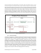
Aminolevulinic acid (ALA) a non-fluorescent non-protein amino acid that is converted into the fluorophore
protoporphyrin IX (PpIX) as part of the heme biosynthesis pathway. The Eagle V1.2 Imaging System’s
fluorescent excitation blue-violet light (405 nm) is maximally absorbed by PpIX fluorophores accumulated
in malignant tissues. The resulting red fluorescence emission (600 – 660 nm) from PpIX in malignant
tissues and green fluorescence emission (500 – 545 nm) from connective tissues and stroma are displayed
in real time on the Handheld Fluorescence Camera’s display screen.
The HFC, Handheld Fluorescence Camera, operates with a number of accessories to ensure specified
performance and safety during use:
i. CSS – Custom Sterile Sleeve
o A sterile sheath to entirely cover the Handheld Fluorescence Camera during use within
the sterile field of an operating room (OR). Refer to Section 2.2.1 and Section 6.3.1 for full
details.
ii. ECH – External Communication Hub
o Mirrors the Handheld Fluorescence Camera’s display on to the display monitor and
facilitates the transfer/export of saved data on the Handheld Fluorescence Camera. Refer
to Section 2.2.2 and Section 6.3.2 for full details.
iii. DIS – Dark Imaging Sheet
o A non-fluorescent plastic sheet providing a consistent and standardized surface
background for Fluorescence Imaging of excised tissues inside the Dark Imaging Box.
Refer to Section 2.2.3 and Section 6.3.3 for full details.
iv. DIB – Dark Imaging Box
o A mechanical assembly providing the optimal dark environment needed to perform
Fluorescence Imaging of excised tissue. Refer to Section 2.2.4 and Section 6.3.4 for full
details.
v. CCD – Contact Charging Device