
Use Instructions

PN 10232 Eagle V1.2 Instructions for Use Rev 1.0 Page 11 of 101
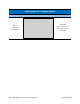
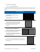
1.4.2.2. Fluorophores – Protoporphyrin IX (PpIX) and Connective Tissue
PpIX
is a fluorophore that accumulates in
cancer cells when a supply of exogenous ALA
is supplied. When excited by 405 nm, PpIX
emits red fluorescent light with a peak
wavelength of approximately 635 nm. This red
fluorescence can be used to visualize and
locat
e the presence of PpIX fluorescent
cancerous tissue. See (Figure 11). In addition,
tissue autofluorescence is also emitted under
405 nm excitation. In breast tissue,
autofluorescence is typically green in color,
largely attributed to autofluorescence of
connective tissues and redox cofactors such as
nicotinamide adenine dinucleotide (NADH)
and flavin adenine dinucleotide (FAD). The
Eagle V1.2 Imaging System captures both the
background green tissue autofluorescence
and the PpIX red fluorescence simultaneously
and in real-time.
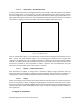
Figure 3 – PpIX Excitation and Emission
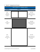
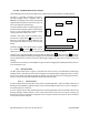
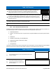
1.4.2.3. Excitation Source – 405 nm Illumination
PpIX has maximum light absorption from blue-violet light at 405 nm (Figure 12). The Eagle V1.2 Imaging
System utilizes safe blue-violet (peak: 405-410 nm) emitting LEDs and an excitation filter to emit narrow
wavelength band centered around 405 nm to induce maximum PpIX fluorescence as well as background
tissue autofluorescence (Figure 4).
Figure 4 – Excitation Light