
Users Manual Part 2

Clinical User’s Guide
IBA | 12-13|
Volume 1 - Treatment Session
|Part II
- Using Treatment Room Equipment Alignment Tools and Devices
|
Working with X-ray Images in a GTR, for Use
With adaPTinsight
Images can be taken either 2D (one image) or 2D stereoscopic (two images that will
be combined).
The adaPTinsight application supports the acquisition of the following kinds of X-ray
images depending on the TR hardware configuration:
Orthogonal X-ray images are taken in the direction orthogonal to the proton
treatment beam.
Portal View X-ray images are taken in the direction of the proton beam.
CBCT X-ray images (3D) using the Rad-B X-ray tube
The Rad-B X-ray tube, which is mounted on the rotating gantry, makes a 180° or
360° rotation around the patient to record a sequence of multiple X-ray images.
A 3D image is then reconstructed by adaPTinsight.
The paragraphs below explain what needs to be done in the GTR prior to acquiring
such X-ray images using adaPTinsight.
Images can be taken in the following mode:
Dual Source mode: both X-ray images can be taken at the same time (i.e., in an
automatic sequence) using two X-ray tubes and two DID panels, and one or two
X-ray generators.
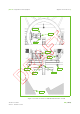
There is one fixed X-ray tube (X-ray tube B) and one retractable X-ray tube (X-ray tube
A), each with an associated flat panel holder system that supports and positions an
appropriate Digital Imaging Device (DID) used to aid in patient positioning. The fixed
X-ray tube is located in the gantry frame 90° counterclockwise from the nozzle. The
retractable X-ray tube is inside the nozzle. The Rad-A X-ray tube is used for portal
view X-ray images and the Rad-B X-ray tube is used for orthogonal X-ray images.
The maximum value of the attenuation equivalent of the couch does not exceed 2 cm
in water (WET).
The flat panel holder systems are mounted on the rotating gantry catwalk behind the
back wall of the patient enclosure.
CAUTION In case of collision of the flat panel with a tough surface or in case of
rough shock, a visual inspection is required to detect any mechanical
deformation. In such case, the use of the detector must be considered
as hazardous and the clinical user shall immediately contact the service
provider.