
Users Manual Part 2


Clinical User’s Guide
IBA | 20-1 |
Volume 1 - Treatment Session
Chapter 20
• • • • • •
Pencil Beam Scanning Principles
Introduction
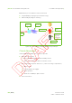
Pencil Beam Scanning (PBS) is the beam delivery technique that delivers the dose by
scanning a narrow proton beam over the target by adjusting the transverse trajectory
of a mono-energetic pencil beam. The scanning operation is performed by two
scanning magnets that are located in the nozzle.
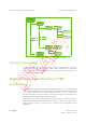
The incoming narrow beam is moved by magnetic scanning in the Xg and Yg
directions (where Xg and Yg are the X and Y axes of the IEC 61217 Gantry Coordinate
System), so as to deflect the beam to the correct position in the tumor. The settings
of the scanning magnets are such that the beam will be positioned at the desired
position and then the beam will be ON up to the moment when the fluence delivered
matches the fluence prescribed for that precise spot in the target in the current
painting or repainting action (this is the ‘spot scanning’ technique, characteristic of
the PBS delivery mode and described in section Spot Scanning.
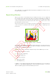
Hence, during a PBS irradiation, the lateral spot position, the beam energy, and the
dose are precisely controlled and adjusted in order for the pencil beam Bragg peak
to cover the patient target volume laterally and in depth and to deliver at each point
of the target the required amount of dose.
The amount of dose delivered at each point of the trajectory is computed by the
Treatment Planning System (TPS) during the dose optimization and calculation
process.
WARNING Only a physicist shall be allowed to attach a fluence file to a patient's
beam.