
User's Manual

Acclarix AX8 Diagnostic Ultrasound System User Manual Imaging
- 47 -
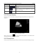
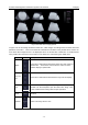
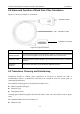
Figure 5-4 3D Image Review
There are two image modes: Volume imaging mode and multi-slice imaging mode. Figure
5-4 shows Volume Imaging Mode in quad screen with a Baby Face volume rendering.
Quadrant A shows a slice through the data that mimics the original ultrasound image.
Quadrant B is orthogonal to A, as if the transducer had been rotated 90 degrees.
Quadrant C is orthogonal to A and B, showing a slice that is parallel to the transducer
face.
The icon at the bottom-right shows the position of each slice with respect to the full 3D
data set.
The following table shows the controls that are available in Volume Imaging Mode
Name
Control
Description
Volume
These two radio buttons toggle between the Volume
display and the Multi-Slice display.
A/B/C/3D
These four radio buttons select which quadrant is the focus
of the navigation/panning controls. A/B/C are three
orthogonal slices through the volume, while „3D‟ is the
rendered image.
Single,
Dual,
Quad
These three radio buttons switch the display to show 1, 2,
or 4 images at once. Single shows the 3D image, dual
shows the A slice and 3D image, Quad shows three MPR
slices and 3D image.
Reset
Reset the operation of pan, rotate and zoom to the initial
condition.
Pan
Pan image along the x-axis or y-axis of the activated
window. Use the trackpad to pan image along x-axis and