
User manual

50
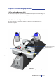
Culture Myograph System Model 202CM
Chapter 7 - Getting Started
7.1 Dissection Protocol for Small Mesenteric Arteries
The culture myograph technique is versatile in that a large variety of physiological
and pharmacological studies of ring preparations from different species can be
performed. Mostly, the culture myograph is used for investigation of small blood
vessels and as an example this chapter describes the dissection of rat mesenteric
arteries.
1. A laboratory rat is euthanized in accordance to the local national law and
regulations. A midline laparotomy is performed to expose the mesenteric bed.
2. Use scissors to remove about 10cm of intestine along with its feeding vas-
culature, including part of the superior mesenteric artery. Be careful not
to damage the vasculature during this procedure. The proximal end of the
intestine section must be about 10cm from pylorus. Make a cut in the proximal
end of the intestine for later identication.
3. Place the excised intestine section in a Petri dish (about 9cm in diameter)
coated with a 5mm thick layer of Sylgard at the bottom to hold the xing pins.
Immediately ll the Petri dish with cold PSS well prebubbled with carbogen (see
Chapter 7.3). The dissection is performed without further oxygenation of the
PSS.
4. Pin down the proximal end of the intestine section on the left-hand side of
the Petri dish without stretching the vessels. Pin down the remaining of the
intestine section in an anti-clockwise direction. In this conguration (proximal
end at the left side, distal end at the right side and running anti-clockwise from
proximal to distal side) the feeding vasculature is on the far side of the intestine
and the veins are usually uppermost.
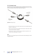
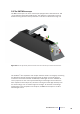
5. Select the vessel segment to be investigated (Fig. 7.1). First time myograph
users are recommended to start dissecting and mounting vessel segments
from the rst or second branch from the superior mesenteric artery
(approximate internal diameter 200-300µm).