
User Manual

ARRHYTHMIA ANALYSIS
PatientNet Operator’s Manual, v1.04, 10001001-00X, Draft 59
All information contained herein is subject to the rights and restrictions on the title page.
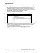
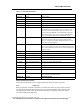
Table 5 1.05 Arrhythmia Analysis
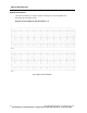
Shape-Related Classification
Shape related arrhythmias occur as single isolated beats and do not form any pattern.
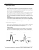
PVC Single PVC
Beats are generally classified as premature ventricular beats if they are early and do not match
the shape of the dominant QRS complex. PVCs are preceded and followed by normal or aber-
rant normal beats. If the criteria for Ventricular Bigeminy or Trigeminy is met, these alarms
are called instead of the single PVC.
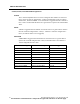
Alarm Label Rhythm Description
MUSCLE Muscle Muscle Artifact
ASYSTOLE Asystole No QRS Detected for 3.0 Sec
CHKSIGNAL Check Signal Intermittent no signal detected
NO SIGNAL No Signal No QRS Detected for 9.0 Sec
V-FIB Ventricular Fibrillation Rapid disorganized Ventricular Impulses, no QRS’s
V-TACH Ventricular Tachycardia Configurable.
The number of consecutive PVCs can be set equal to, and
between, 3 and 8. The Heart Rate can be set equal to, and
between, 100 and 120 BPM. A V-TACH alarm is triggered
when the consecutive PVC count is reached AND the heart
rate is greater than or equal to the set Heart Rate Value.
V-RUN Ventricular Run V-RUN is triggered when the number of consecutive PVCs is
greater than 2 and less than the V-TACH configured PVC
value (i.e. when the V-TACH is configured at 3 PVCs, the V-
RUN alarm is never triggered).
V-RHYTHM Ventricular Rhythm V-RHYTHM is triggered when the number of consecutive
PVCs is greater than or equal to the V-TACH configured PVC
value, but the Heart Rate is less than the V-TACH configured
heart rate value.
BIGEMINY Ventricular Bigeminy N-PVC-N-PVC-N-PVC Sequence
TRIGEMINY Ventricular Trigeminy N-N-PVC-N-N-PVC-N-N-PVC Sequence
COUPLET Ventricular Couplet 2 Consecutive PVCs
HIGH PVC High PVC PVC Count > High PVC Limit
PVC PVC Single PVC
SV-TACH Supraventricular Tachycar-
dia
8 Consecutive SVEs, HR 150 or More
HIGH HR High Heart Rate HR Greater than High Rate Limit
LOW HR Low Hear Rate HR less than Low Rate Limit
NO ARR Arrhythmia unable to ana-
lyze
No good leads are available for analysis
CHK LEAD Check Lead One or more of the ECG leads has a poor connection and/or is
causing significant baseline wander
REGULAR Normal Dominant Rhythm