
User Manual


SKIN PREPARATION AND LEAD PLACEMENT
PatientNet Operator’s Manual, v1.04, 10001001-00X, Draft 49
All information contained herein is subject to the rights and restrictions on the title page.
Patients with a Pacemaker
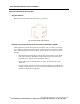
Note: Do not place an electrode near the implant. Move the relevant electrode at
least four inches, either lower on the chest or near the scapula. The chest
lead may also have to be moved.
Note: Initiate the learning process when the patient is in their dominant rhythm.
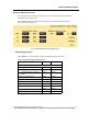
Fig. 16. Pacemaker Patients Lead Placement
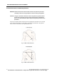
The transceiver detects pacemaker pulses on Leads I and II. The more parallel these
vectors are to the pacing vectors, the more likely the pacer pulses will be detected. The
impedance pulse vector is typically positioned between the pacemaker implant and the
pacing lead in the bottom of the heart. In adults, the pacemaker is usually implanted in
the left or right pectoral area. To better reject the impedance pulses, the following lead
placement guidelines may help.
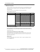
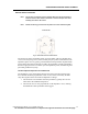
Left Arm Implant: Reposition the LA Electrode
The impedance vector runs from the left pectoral region to the bottom of the heart.
Since Lead II should be perpendicular to this vector, Lead I is most likely the diffi-
culty. Moving the left arm electrode accomplishes two things:
1. The electrode is moved farther from the pacemaker; typically three or four
inches below the standard lead position.
2. The Lead II vector is more perpendicular to the impedance vector, reducing
the likelihood of false pacemaker detect triggers.