
User guide


Charnwood Dynamics Ltd. Coda cx1 User Guide - Gait Analysis II - 1
CX1 USER GUIDE - COMPLETE.doc 26/04/04
66/162
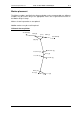
The sides of the Pelvic frame should be tilted so that the plane defined by the ‘PSIS’ and
‘ASIS’ markers includes the Pelvis ASISs and PSISs.
Firstly, the line joining the ‘L.ASIS’ and ‘R.ASIS’ markers defines the medio-lateral axis of
the Pelvis. The anterior-posterior axis direction is then defined by the perpendicular to the
ASIS - ASIS line in the plane which contains all three Pelvic frame markers.
‘R.Front.Wand’ & : Optional markers on a forward wand, in-line with the sacral wand.
‘L.Front.Wand’ If present, this marker allows the pelvis orientation to be tracked
more accurately when one of the other Pelvic frame markers is
obscured (e.g. by swinging arm), or when the sacral wand marker is
out of the field of view.
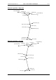
Leg and Foot Markers
The knee, ankle, heel, toe and (optional) hip markers are placed directly on the skin. The
remaining leg markers are mounted at both ends of specially hinged femoral and tibial
wands. The tibial wand incorporates extra bracing against the rigid, bony, middle region
of the shank. The femoral wand should be strapped to the thigh just above the knee but
below the major bulk of thigh muscle. Both wands are to be hinged on the lateral aspect
of the leg; the hinge allows for easy adjustment after the wand is fitted. The exact
positioning of the wands is not critical except for the (local transverse projection of the)
orientation which must be perpendicular to the knee-joint axis in the case of the femoral
wand and, for the tibial wand, perpendicular to the ankle-joint axis. The wands thereby
define the orientations of the segment local embedded (transverse) axes, but do not
correspond to any particular anatomical landmarks.
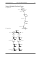
Unilateral data acquisition:
‘Ant.Fem.’ : Positioned, facing laterally, at the front of the femoral wand.
‘Post.Fem.’ : Positioned, facing laterally, at the rear of the femoral wand.
The femoral wand should be rotated at the hinge until perpendicular to the knee axis.
‘Ant.Tib.’ : Positioned, facing laterally, at the front of the tibial wand.
‘Post.Tib.’ : Positioned, facing laterally, at the rear of the tibial wand.
The tibial wand should be rotated at the hinge until perpendicular to the ankle axis.
‘Ankle’ : Positioned on the lateral aspect of the medio-lateral ankle axis.
‘Heel’ : Positioned, facing laterally, at the heel.
‘Toe’ : Positioned, facing laterally, at the end of the 5
th
metatarsal.
‘Knee’ : Positioned on the lateral aspect of the medio-lateral knee axis.
[‘Hip’] - optional : Positioned on the lateral aspect of the medio-lateral hip axis
Even if present, the ‘Hip’ marker plays no role in the segment model and, in particular, has
no influence whatsoever on the calculated positions of hip joint centres which are derived
entirely from the goemetry of the pelvis.
Bilateral data acquisition:
Both sides as above but NOTE: the marker names are preceded by “L.” or “R.” (e.g.
‘L.Toe’).