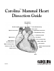
TM Carolina Mammal Heart Dissection Guide Left common carotid artery Brachiocephalic artery Superior vena cava Left subclavian artery Aortic arch Ligamentum arteriosum Left pulmonary artery Pulmonary trunk Right auricle Left atrium Left coronary artery Right atrium Coronary sulcus Great cardiac vein Left ventricle Right coronary artery Anterior cardiac vein Anterior interventricular sulcus Right ventricle Apex C80136
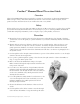
CarolinaTM Mammal Heart Dissection Guide Overview The Carolina Mammal Heart Dissection Guide is a general set of instructions for dissecting mammal hearts. With each type of heart, there will be differences in the size of the structures and heart regions, but the general structures and their relative location will be the same or very similar. Safety Follow safe laboratory practices when performing any dissection. Wear safety glasses or goggles, gloves, and lab aprons when dissecting.
10. Refer to the dissected mammal heart image again. Make an incision through the left ventricle inferior to the interventricular groove. Remove the lower front portion of the wall. Observe the size of the left ventricle in relation to the right ventricle. Observe the muscular interventricular septum that divides the two chambers. 11. Observe the bicuspid valve supported by chordae tendinae and papillary muscles. 12.
Carolina’s Perfect Solution® Independent, certified laboratory analyses of specimens fixed in Carolina’s Perfect Solution® have found it to be nontoxic and free of dangerous off-gassing. This means that, for safety purposes, classrooms and labs using Carolina’s Perfect Solution specimens do not require specialized ventilation. Carolina does recommend using some active ventilation when working with any preserved specimens or chemicals.