Bio-Dot® SF Microfiltration Apparatus Instruction Manual Catalog Number 170-6542 170-6543 For technical service call your local Bio-Rad office or in the U.S., call 1-800-424-6723.
Table of Contents Page Section 1 Introduction ..................................................................................1 1.1 Specifications............................................................................................1 Section 2 Special Handling and Features ..................................................1 2.1 2.2 Autoclaving ..............................................................................................1 Chemical Stability ....................................
Section 1 Introduction The Bio-Dot SF blotting apparatus has an evenly spaced, slot shaped sample template for easy slot blot sample comparisons. Because the Bio-Dot SF apparatus focuses the applied samples in a thin line instead of a circle, this slot format makes it easy to use a densitometer to quantitate results. The Bio-Dot SF apparatus is provided as a complete unit, or as a modular addition to the Bio-Dot microfiltration system.
Section 2 Special Handling and Features The Bio-Dot apparatus withstands autoclave temperatures for sterilization, as well as cleaning with alcohols, acids, and base solutions. 2.1 Autoclaving The Tygon tubing and flow valve cannot be autoclaved. All other components of the apparatus withstand the autoclave treatment. After repeated autoclaving (~25 cycles) the silicone rubber gasket may need replacing.
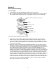
Section 3 Bio-Dot SF Assembly 3.1 Assembly 1. Clean and dry the Bio-Dot SF apparatus and gasket prior to assembly. 2. Place the gasket support plate into position in the vacuum manifold. (There is only one way to slide the plate into the manifold.) Place the sealing gasket on top of the vacuum manifold. Sample template with attached sealing screws Membrane Filter paper (3 sheets) Sealing gasket Gasket support plate Vacuum manifold Tubing and flow valve Fig.1.
5. Place the sample template on top of the membrane. The guide pins ensure that the template will be properly aligned. Finger-tighten the four screws. When tightening the screws, use a diagonal crossing pattern to ensure uniform application of pressure on the membrane surface (see Figure 2). Fig. 2. Diagonal crossing pattern for tightening screws in the Bio-Dot apparatus. 6.
3.2 Helpful Hints 1. During the assay, do not leave the vacuum on. This may dehydrate the membrane and may cause halos around the wells. Apply vacuum only until solutions are removed from the sample wells, then adjust the flow valve so that the unit is exposed to air and disconnect the vacuum source. 2. If some sample wells are not used in a particular assay, those wells must be closed off to insure proper vacuum to the wells in use. There are three ways to close off unused wells.
Section 4 Protein Slot Blotting 4.1 General Recommendations 1. Solution Volume. The liquid in the incubation vessel should be least 0.25 cm deep to ensure the membrane is completely submerged during incubation. There should be at least 0.5 ml of reagent per cm2 of membrane. Larger volumes may be used for convenience. 2. Handling the mMembrane. Wear clean plastic gloves or use forceps to avoid fingerprints on the membrane. 3. Temperature. All steps are performed at room temperature (22–25°C). 4.
5. Wash each sample well with at least 200 µl of TBS. Pull the wash liquid through by applying gentle vacuum (flow valve setting 3, Figure 3). 6. After the wells have completely drained, remove the membrane from the apparatus. The best method for removing the membrane from the Bio-Dot SF apparatus is to leave the vacuum on following the wash step. With the vacuum applied, loosen the screws and remove the sample template. Next, turn off the vacuum and remove the membrane. 7.
Section 5 DNA Slot Blotting This section gives protocols for DNA slot blotting. The alkaline blotting method, using Zeta-Probe membrane, and a more standard method for DNA blotting to nitrocellulose, are described. 1. The target DNA must be denatured prior to application to the membrane. When using the Zeta-Probe membrane, denature the DNA sample by addition of NaOH and EDTA solution to final concentrations of 0.4 M NaOH, 10 mM EDTA. Heat the sample to 100°C for 10 minutes to ensure complete denaturation.
Section 6 RNA Slot Blotting RNA must be denatured prior to application to Zeta-Probe® or nitrocellulose membranes to ensure optimal hybridization. Two protocols are presented for denaturing RNA samples. 6.1 Alkaline RNA Denaturation and Fixation 1. Always wear gloves when handling blotting membranes. Prewet the blotting membrane by placing it gently at a 45° angle into a tray of wetting solution.
Note: A method for applying gentle vacuum to the apparatus is to adjust the flow valve to setting 3. Use a finger to cover the valve port exposed to air. The amount of vacuum reaching the manifold will be regulated by the pressure of your finger on the valve. 6. Rinse all wells to wash any sample on the side of the wells through. Rinse with 500 µl TE. Apply vacuum (flow valve setting 1, Figure 3) until the sample wells are dry. 7. Disassemble the Bio-Dot SF apparatus. Remove the blotted membrane. 8.
Section 7 Hybridization Protocols for Nucleic Acids 7.1 Probe Recommendations The specific activity, concentration, size range, and purity of the probe all have an important effect on signal-to-noise ratio during hybridization.
The carrier DNA used with nitrocellulose must be denatured before adding it to the prehybridization solution. Heat the DNA at 100°C for 5 minutes and cool rapidly. 3. Seal the top of the bag and incubate. For DNA or RNA Bound to Zeta-Probe Membrane For DNA Bound to Nitrocellulose For RNA Bound to Nitrocellulose 5 minutes at 65°C 2–4 hours at 68°C 8–20 hours at 42°C Hybridization 1. Cut one corner of the plastic bag.
2. After washing, the blotted membrane is ready for autoradiography. If no further cycles of hybridization are to be done on the membrane, the membrane can be dried. When reprobing, do not allow the membrane to dry between hybridizations. Make the autoradiographic exposure with the moist membrane wrapped in plastic wrap or enclosed in a sealable plastic bag.
Washes 1. At the completion of hybridization, remove the membranes from their hybridization bags into 2x SSC. Rinse briefly, then wash them sequentially with agitation for 15 minutes at room temperature in the following solutions: • 2x SSC/0.1% SDS • 0.5x SSC/0.1% SDS • 0.1x SSC/0.1% SDS 2. For DNA bound to nitrocellulose membranes, it may be necessary to include an RNase treatment in the wash. Membranes are treated with 20 µg/ml RNase for 30 minutes at 37°C in 2x SSC (Johnson et al. 1984). 3.
Section 8 Solutions for Protein Applications 8.1 Solutions for Nitrocellulose Membrane Tris-Buffered Saline, 1x TBS, 2 L 20 mM Tris, pH 7.5 500 mM NaCl Dissolve 4.84 g Tris, 58.48 g NaCl in ddH2O. Adjust to pH 7.5 with HCl. Adjust the volume to 2 L with ddH2O. Tween 20 Wash Solution, 1x TTBS, 1 L 20 mM Tris, pH 7.5 500 mM NaCl 0.05% Tween 20 Add 0.5 ml Tween 20 to 1 L of TBS. Blocking Solution, 100 ml 3% gelatin-TBS Add 3.0 g gelatin to 100 ml TBS. Heat at 37°C to dissolve the gelatin.
8.2 Solutions for Zeta-Probe Membrane Two methods of blocking are given. Method A uses nonfat dry milk as the blocking agent. Method B uses gelatin and MPO** as the blocking agents. The solutions for the two methods are not interchangeable. If Method A is chosen, all solutions must be prepared according to Method A; if Method B is chosen, all solutions must be prepared according to Method B. TBS, Tris buffered saline, 2 L Same as nitrocellulose membrane solution.
Section 9 Solutions for Nucleic Acid Applications For DNA or RNA Bound to Zeta-Probe Membrane For DNA Bound to Nitrocellulose For RNA Bound to Nitrocellulose 1 mM EDTA 7% SDS 0.5 M NaHPO4, pH 7.2 6x SSC 0.5% SDS 5x Denhardt’s solution 100 µg/ml denatured salmon sperm DNA 1 mM EDTA 50% formamide 5x SSC 1x Denhardt’s solution 50 mM NaHPO4, pH 6.5 250 µg/ml denatured salmon sperm DNA 20x SSC 3 M NaCl 0.3 M trisodium citrate (FW = 294.1) Dissolve 175.0 g NaCl and 88.2 g trisodium citrate in ddH2O.
50% Formamide Dilute 50.0 g formamide to 100 ml with ddH2O. Store at 4°C. Immediately before use, deionize the required volume by stirring gently for 1 hour with 1 g mixed bed ion exchange resin (AG® 501-X8 (D) resin, catalog number 142-6425)/10 ml of formamide. Filter through coarse filter paper. For DNA or RNA Bound to Zeta-Probe Membrane For DNA Bound to Nitrocellulose For RNA Bound to Nitrocellulose A. Wash 2 times for 30–60 minutes at 65°C in: 1 mM EDTA 40 mM NaHPO4, pH 7.2 5% SDS A.
Section 10 Troubleshooting Guide I. Filter Apparatus 1. Leakage or cross-well contamination a. Improper assembly. The screws must be retightened under vacuum following the initial assembly. The thickness of filter paper must be correct, or leakage will result. Exactly three sheets of filter paper must be placed on the membrane support. Do not use filter paper other than the Bio-Dot SF filter paper. b. Membrane is not properly rehydrated after assembly.
III. High Background After Incubation with Labeled Probes 1. DNA and RNA a. Unincorporated label, small radioactive decay products, and small probe fragments resulting from nick-translation can increase overall background. Use the Bio-Spin® chromatography columns to remove unincorporated label. Filter hybridization solutions before use. Use the probe as soon as possible after preparation. Reduce exposure of the probe to DNase during nick translation. b. Improper blocking conditions were used.
b. Hybridization was insufficient. Incorporate 10% dextran sulfate in the hybridization mixture. This polymer effectively reduces the solvent volume, thereby increasing the concentration of the solutes and enhancing hybridization. c. Exposure time was insufficient. Increase the time of exposure. d. Sample load was insufficient. Increase the sample load. e. Probe concentration is too low. If low signal is accompanied by low background, then the probe concentration can be increased. f.
Section 11 References Achberger EC and Whiteley HR, The role of the delta peptide of the Bacillus subtilis RNA polymerase in promoter selection, J Biol Chem 256, 7424–7432 (1981) Allen JD and Parsons SM, Nitrocellulose filter binding: quantitation of the histidyl-tRNA-ATP phosphoribosyltransferase complex, Anal Biochem 92, 22–30 (1979) Bennett FC and Yeoman LC, An improved procedure for the 'dot immunobinding' analysis of hybridoma supernatants, J Immunol Methods 61, 201–207 (1983) Berg LJ et al.
Karagyozov LK and Hadjiolov AA, Isolation of active transcription complexes from animal cell nuclei by nitrocellulose binding, J Biochem Biophys Methods 5, 329–339 (1982) Kranz RG and Gennis RB, A quantitative radioimmunological screening method for specific gene products, Anal Biochem 127, 247–257 (1982) Kutateladze TV et al.
Section 12 Ordering Information Catalog # Description 170-6542 Bio-Dot SF Apparatus 170-6543 Bio-Dot SF Module, for Bio-Dot to Bio-Dot SF conversion 170-6544 Bio-Dot SF Gaskets, 2 162-0161 Bio-Dot/Bio-Dot SF Filter Paper, 11.3 x 7.7 cm, 60 sheets 170-6545 Bio-Dot Apparatus 170-6546 Bio-Dot Gaskets, 3 170-6547 Bio-Dot Module, for Bio-Dot SF to Bio-Dot conversion 162-0117 Nitrocellulose Membranes, 0.
Bio-Rad Laboratories, Inc. Web site www.bio-rad.