
User`s guide

Imaging Modalities
Tissue Doppler Imaging
Revision D.0 7-123
ssn February 10, 1999 C:\WINNT\Profiles\dapowell\Desktop\D.0 Books\CD FILES SONOS
D.0\System Basics D.0\Frame Files\7CH.FM add.2
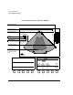
Using Tissue Doppler Imaging
The following section describes Tissue Doppler controls and how to use them.
Note the following Doppler mode guidelines:
•Touching PW or CW turns on Spectral, unless depth marks are used.
•In PW nonspectral mode, pressing the key automatically turns on
Spectral.
• Spectral is available in 2D/BMode, Color, and Angio when depth marks
are used.
•In Spectral, pressing turns on BMode Live (2D-Live if in Cardiac).
• When PW is turned on, the gate automatically moves away from the edge of
the image.
•Touching Cursor Angle when highlighted sets the angle to zero, except
when Intelligent Doppler is active or the image is frozen.
•In PW Spectral, you can display Heart Rate from a Doppler trace and
make a measurement.
For more information on PW and CW modes, see “PW and CW Imaging” on
page 7-79.
Depending on the imaging mode being used, the Tissue Doppler controls
include
•Color Gain
• Smoothing
•Color Map
• B/W Suppress
• PW Scale
• Reject
•Colorize
•Gate Length
•Compress
•Power
Enter
Enter