
User`s guide


Imaging Modalities
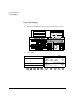
PW and CW Imaging
Revision D.0 7-105
ssn February 10, 1999 C:\WINNT\Profiles\dapowell\Desktop\D.0 Books\CD FILES SONOS
D.0\System Basics D.0\Frame Files\7CH.FM add.2
PW and CW Troubleshooting
Always make sure the active preset is appropriate for the study being performed.
If necessary, adjust the and
monitor controls for the ambient light.
Symptoms Suggestions
Not sensitive. Power. Gain, Compress, and Reject
to increase the amount of Doppler information
displayed.
For cardiac images, reposition the transducer to make
the beam parallel to flow.
For vascular images, use Cursor Angle to obtain the
optimal angle to flow. Position the cursor parallel to
and in the same direction as blood flow. To change
your preferred angle to flow, press and adjust
Optimum Angle.
Change to a lower frequency transducer.
Try using Colorize, to improve contrast resolution.
PW Only:
Gate Length to increase the sample volume size.
CW Only:
Place the diamond (focal point) on the cursor line over
the area where the greatest sensitivity is needed.
2D/BMode
reference image
does not update.
Make sure 2D Hold or BMode Hold is turned off.
If an R-wave is present Delay or Beats and
Interval (all Physio Trigger controls). For
systems without physios, make sure Duplex is off,
press , and adjust Interval.
Check ECG leads for proper placement.
ECG Gain to ensure triggering.
Difficult to get a
good acoustic
window.
Try using a nonimaging transducer.
Setup
Setup