
User`s guide

Imaging Modalities
PW and CW Imaging
Revision D.0 7-79
ssn February 10, 1999 C:\WINNT\Profiles\dapowell\Desktop\D.0 Books\CD FILES SONOS
D.0\System Basics D.0\Frame Files\7CH.FM add.2
PW and CW Imaging
Introduction
Pulsed Wave Doppler (PW) mode provides a “spectral” representation of the
velocity of blood flow or tissue movement. Velocities are represented on graphs
that use a cm/sec scale to enable velocities to be measured and tracked over time.
The “Pulsed” in PW means that the transducer is sending the ultrasound pulse
and receiving its echo through a single crystal. This permits you to sample
velocities at specific depths.
Continuous Wave Doppler (CW) provides the same spectral display as PW, but
the transducer uses separate crystals to transmit ultrasound pulses and receive
their echoes. This permits you to sample higher velocities than in PW mode. But
CW samples along the entire line of sight, and can exhibit more interference from
movement.
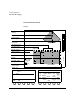
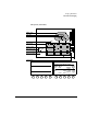
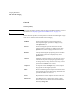
This section describes
• PW and CW screens and touch panels (page 7-80)
• Controls (page 7-82)
• Using PW imaging (page 7-93)
• Using CW imaging (page 7-101)
• Troubleshooting PW and CW images (page 7-105)