
User manual


171
However, if 2 slices were obtained from 1 animal, and these two slices were exposed to the SAME
stimulation protocol and the SAME perfusion solutions, some would consider this to be an N of 2, and
others would consider it a ‘Strict’ N of 1.
In general we at WinLTP Ltd. do not favor exposing multiple slices obtained from 1 animal to the SAME
stimulation protocol and the SAME perfusion solutions, and WinLTP was not designed for this approach.
WinLTP was designed to provide, if necessary, completely DIFFERENT stimulation protocols and
perfusion solutions. Four instances of WinLTP can be run on one computer, and each of these WinLTPs
can control completely separate stimulation and perfusion solutions. And taking 4 slices from 1 animal
and exposing them to DIFFERENT stimulation and perfusion solutions is clearly more difficult to do than
taking 4 slices from 1 animal and exposing them to the SAME stimulation and perfusion solutions.
That said, if your experiment includes testing DIFFERENT concentrations of agonist or antagonist, then
running several slices obtained from 1 animal and exposing these slices to the SAME stimulation protocol
but DIFFERENT perfusion solutions, this would be an easy way to run many slices side by side, have N
equal to the number of slices tested, and yet change all the solutions easily and manually without
requiring automated perfusion control.
WinLTP just tries to provide the tools for all situations, including completely independent control of
stimulation and perfusion solutions.
10.2.3 Automated Perfusion Problems – Dead Volume in Extracellular Slice
Experiments
Before we go into setting up the automated perfusion control, it is important to discuss what we see as
potentially the main problem of automatic perfusion control in extracellular slice experiments, namely
dead volume.
For extracellular slice experiments, when aerated physiological saline has stayed in polyethylene or tygon
tubing for a ‘long’ period of time before being perfused on slices, oxygen can diffuse across the tubing
wall. In addition this loss of oxygen/carbon dioxide, can also cause changes in pH if a standard
bicarbonate buffer is used to perfuse the slice.
We honestly don’t know how bad this lack of oxygen and pH change is, but it depends on the thickness of
the tubing wall, what the tubing is made of, the volume of the solution that just ‘sits’ there before being
perfused onto the slice (the dead volume), and how long it ‘sits’ there. More on how to reduce this dead
volume is presented below.
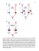
For extracellular slice experiments, the important dead volume for the standard perfusion system is
between the aerated perfusion bottle and the manifold (shown in red in Fig 10.2.4.1A), and for the pre-
flush perfusion system between the valve and the manifold (shown in red in Fig 10.2.4.1B).
Estimates of dead volume for 1.32” and 1.00mm inner diameter tubing for the pre-flush system (only
between the T-fitting, in the valve, and to the manifold) for 9cm (for a 4 channel system, and 16cm (for an